HK3 - Thermal neutron analytical methods
Three instruments HC3-a HC3-b and HC3-c operate at the thermal neutron beam formed by short neutron guide tube. The neutron guide ensures an efficient transport of thermal neutrons from a Ć 100 mm horizontal channel of the reactor to small target areas of the HC3-a, HC3-b and HC3-c spectrometers. The neutron guide is built of a mirror type tube of rectangular cross-section, cylindrically bent in the vertical direction. It consists of 15 mirror sections made of glass plates of float type. The surface of these plates is coated with Ni reflecting layer with thickness of 2000 Å. The internal cross-section of each mirror section is 4×150 mm2. The overall length of the guide is 5.63 m, the curvature radius being 825 m. In order to suppress a background due to a direct beam of gamma rays and fast neutrons, the guide is tightly surrounded by a combined shielding consisting of lead and polyethylene pellets. Unlike thermal neutrons, gamma rays and fast neutrons are not subject to reflections from the Ni coating and penetrate the guide walls. In the shielding around they are scattered and absorbed and only collimated thermal neutrons pass through. In addition, a biological shielding, formed by boron-doped polyethylene and lead bricks, is built along the whole guide. The shape of the incoming neutron beam at the entrance of the guide matches the cross-section of the guide. This has been achieved using a 90 cm long collimator made of lead with a rectangular aperture. Immediately behind the guide exit, the neutron beam is tailored by an additional collimator made of 6Li2CO3 to reduce the beam cross-section to 4×60 mm2. The flux of thermal neutrons at the guide exit averaged over the beam cross-section is (1.5±0.2)·107n cm-2 s-1. The cadmium ratio is equal approximately to 105. The above mentioned fluxes refer to the reactor power of 8 MW. Beyond the guide exit, the vertical divergence of neutron trajectories is characterized by angular deviations below 0.5°.
Video: Neutron Depth Profiling for Li-Ion Batteries
Video-clip on the use of nuclear-analytical technique for research and development mainly in the field of novel batteries.
Produced in the frame of NPI participation in SINE2020 project (HORIZON 2020 grant agreement No. 654000), workpackage "Industry consultancy“.
Managed by: Neutron Physics Laboratory (NPL) of CANAM infrastructure, Nuclear Pfysics Institute, Czech Academy of Sciences. Made by: Maurfilm.
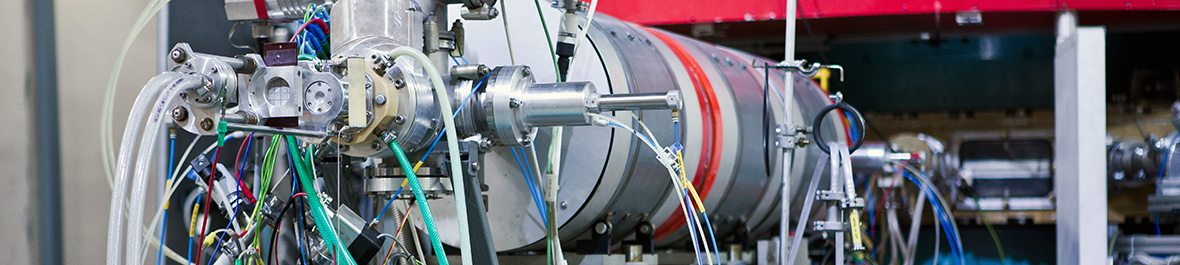
Thermal Neutron Depth Profiling (NDP) facility was set up just behind the neutron guide at HK3. The multidetector spectrometer consists of a large vacuum chamber, automatic target holders and several different data acquisition systems which can be used at the same time. NDP is the nuclear analytical technique available to profile light elements in solids. It utilizes the existence of isotopes of elements that produce prompt monoenergetic charged particles upon capture of thermal neutrons.
From the energy loss spectra of emitted products the depth distributions of light elements can be reconstructed. The NDP method is an excellent tool for studies of numerous problems in solid-state physics (diffusion, sputtering), material science (corrosion), electronics, optronics, life sciences etc. Its applicability and efficiency has steadily expanded.

List of nuclides exploited in NDP analyses
| Nuclide | Natural abundance or activity |
Nuclear reaction | Energy of reaction products | Cross section | Detection limit | |
| [atoms/mCi] % | E1 [keV] | E2 [eV] | [barns] | [at/cm2] | ||
| 3He | 0.00013 | 3He(n,p)3H | 573 | 191 | 5326 | 3.1·1013 |
| 6Li | 7.42 | 6Li(n,a)3H | 2051 | 2734 | 94 | 1.8·1014 |
| 7Be* | 2.5·1014 | 7Be(n,p)7Li | 143 | 207 | 48000 | 3.5·1012 |
| 10B | 19.6 | 10B(n,ag)7Li | 1471 | 839 | 3606 | 4.3·1013 |
| 10B | 19.6 | 10B(n,a)7Li | 1775 | 1014 | 230 | 6.7·1014 |
| 14N | 99.64 | 14N(n,p)14C | 584 | 42 | 1.81 | 9.1·1016 |
| 17O | 0.037 | 17O(n,a)14C | 1415 | 404 | 0.24 | 7.1·1017 |
| 22Na* | 4.4·1015 | 22Na(n,p)22Ne | 2247 | 103 | 31000 | 4.7·1012 |
| 33S | 0.76 | 33S(n,a)30Si | 3091 | 412 | 0.14 | 1.2·1018 |
| 35Cl | 75.5 | 35Cl(n,p)35S | 598 | 17 | 0.49 | 3.4·1017 |
| 40K | 0.012 | 40K(n,p)40Ar | 2231 | 56 | 4.4 | 3.8·1016 |
| 59Ni* | 1.3·1020 | 59Ni(n,a)56Fe | 4757 | 340 | 12.3 | 1.4·1016 |
| 209Bi | 100.0 | 209Bi + n -> 210Bi | 0.02 | 1.2·1019 | ||
| 210Bi -> b + 210Po | ||||||
| 210Po -> a + 206Pb | 5300 | |||||
Instrument Parameters
| Beam cross-section | 4 x 60 mm |
| Neutron flux | 1·107 n/cm2s |
| Sample size | 50 - 1000 mm 2 |
| Detector systems: | |
| - standard arrangement (single detector facing the sample) |
4 |
| - sandwich arrangement large angle coincidence spectroscopy (2-dimensional data processing) |
1 |
| - dE-E telescope arrangement | 1 |
| - pulse shape discrimination analysis | 2 |
| Solid angle of the detector-sample system | 0.001% - 0.1% |
| Detectors | PIN photodiode - HAMAMATSU Charged particle detector - CANBERRA |
| Detector areas | 50 - 300 mm |
| Depletion depth | 10 - 100 µm |
| Detection limit | |
| - standard arrangement - N, Cl | 10-3 % at. |
| - He, Li, B | 10-5 % at. |
| - sandwich arrangement - Li, B | 10-8 % at. |
| Depth resolution | 10 - 50 nm |
| Maximum detectable depth interval | 3 - 70 µm |
 Spectrometer NDP
The facility for the measurements of 10B concentrations in biological samples includes HPGe detector with 25% relative efficiency and associated Pb - 6Li2CO3 shielding. Described facility is installed at the distance of 1 m from the exit of the neutron guide. At the target position the neutron flux is approximately 3×10⁶ n cm-2 s-1 at the reactor power of 8 MW. This facility makes it possible to determine 10B concentration of 1 ppm in 1 ml samples with statistical uncertainty of 5% within 15 min. This instrument is in a common property with Nuclear Research Institute, plc.
Applications
The experimental set-up was mainly developed for the on-line determination of 10B concentrations of ppm-order in biological samples. This facility can also be used for determination of other isotopes with a sufficiently large (n,g) cross-section.
Instrument Parameters
| Neutron flux | 3·106 n/cm-2 s-1 | |
|
Beam cross section at target position
|
25 x 5 mm2 | |
|
Detector
|
HPGe (25% relative efficiency) | |
| Sample enclosure for liquid and powder sample | Teflon cylinder vial: Diameter - 10mm & Height - 10mm |
Expected interference-free detection limit for PGAA instrument at research reactor LVR-15 assuming 24h irradiation.
| Element | Det. limit (µg) | Eγ (keV) | Element | Det. limit (µg) | Eγ (keV) |
| Hydrogen | 20 | 2223 | Cobalt | 20 | 230, 556 |
| Boron | 0.06 | 478 | Nickel | 200 | 283, 465 |
| Nitrogen | 4000 | 1885, 5298 | Copper | 10 | 159, 278 |
| Sodium | 70 | 472, 869 | Zinc | 700 | 115, 1077 |
| Magnesium | 2000 | 585,1809 | Selenium | 40 | 239 |
| Aluminium | 500 | 1779, 7724 | Molybdenum | 150 | 720, 778 |
| Phosphorus | 2000 | 637, 1072 | Silver | 30 | 192, 236 |
| Sulfur | 300 | 840, 2379 | Cadmium | 0.1 | 559, 651 |
| Chlorine | 10 | 517, 786 | Samarium | 0.03 | 333, 439 |
| Potassium | 100 | 770, 7771 | Gadolinium | 0.02 | 182, 1186 |
| Calcium | 600 | 519, 1943 | Gold | 30 | 215 |
| Titanium | 40 | 342, 1381 | Mercury | 1.5 | 368 |
| Chromium | 150 | 749,834 | Lead | 40000 | 7368 |
| Manganese | 30 | 847, 1811 | Neodymium | 10 | 619, 697 |
| Iron | 300 | 352, 7631 | Indium | 5 | 162, 186 |
Application Example
The deterimantion of boron concentration in graphene powder.
Electronic properties of graphene can be changed by doping with electron-donating (nitrogen, phosphorus) or electron-withdrawing (boron) groups. Level of doping (concentration of doped group) is crucial for for tuning the electrochemistry performance of graphene. Boron-doped were prepared by thermal exfoliation of graphite oxide in an atmosphere with boron trifuoride diethyl etherate at high temperatures. The effect of exfoliation temperature as well as of hydrogen content in atmosphere were investigated using PGAA at LVR-15 Řež.
Special software was developed for precise fitting of Doppler-broadened boron peak at 478 keV.
Fig. 1 Example of Doppler-broadened boron peak at 478 keV in gamma spectrum of graphene powder.
Two-germanium-detector system is employed for the study of γ-γ coincidences from (n,g) reactions. The horizontally oriented detectors are placed above and bellow the horizontally oriented target, their axes being mutually parallel. At present, the bottom detector is of the HPGe type with the 20% relative efficiency and the energy resolution of 1.9 keV at Eg= 332 keV, while the Ge(Li) detector with the 12% relative efficiency and 2.1 keV energy resolution at 1332 keV is located above the target. These detectors can be easily replaced by another ones. The distance between cylindrical surfaces of the germanium crystals is 4 cm only. The neutron flux at the target position is (2.8 ± 0.5)·106 n cm-2 s-1 at the reactor power of 8 MW. The coincidence efficiency for a g-cascade from the 60Co source is 3·10 -5, including the effect of the solid angle. The detectors are shielded from g-ray and neutron background in the reactor hall by a combined shielding. The electronic system is based on the standard fast/slow coincidence arrangement. The events are accumulated by a PC computer.

Applications
The facility was designed primarily for the study of photon–strength functions, but it can also be used for nuclear structure studies via g-g coincidences following the thermal neutron capture.
Instrument Parameters
|
Neutron flux at the target position
|
3·106 n/cm2 s |
|
Cadmium ratio
|
105 |
|
Beam cross section at target position
|
25 x 5 mm2 |
| Detectors | HPGe (20% relative efficiency) Ge(Li) (12% relative efficiency) |
| Coincidence full-energy-peak efficiency | 3·105 for 60Co (1173 keV+1332 keV) |