Research
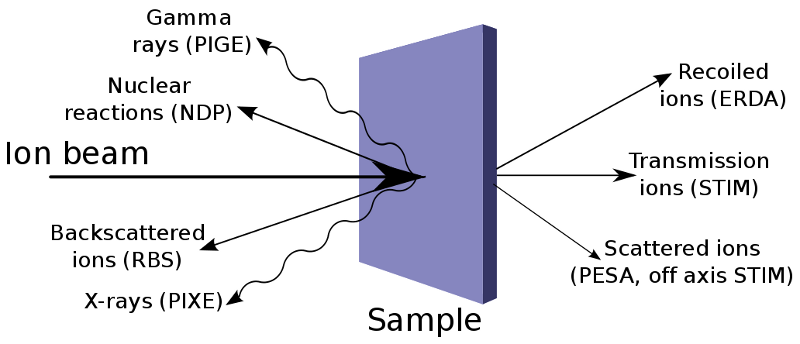
Ion beam analytical methods is a cluster of analytical techniques that use an ion beam for study of structures and composition of samples. The beam of the accelerated ions is applied for the surface modification of solids and for the analysis of their composition and structure. Ion beams are produced by a particle accelerator. The individual IBA techniques are distinguishable by the different types of interaction of ion beam and solid target. These methods have several unique characteristics which couldn't be substituted for other alternative approaches.
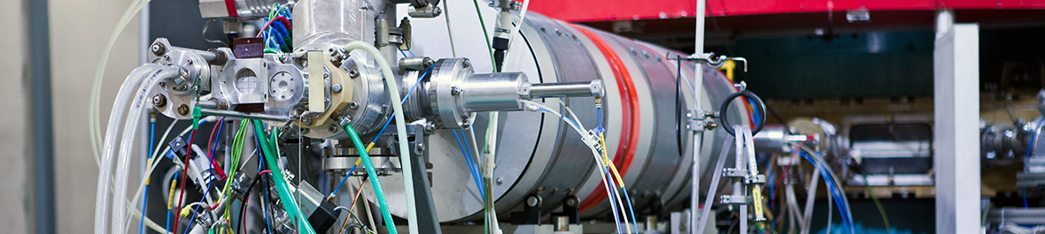
In department we use 3 MV electrostatic accelerator Tandetron 4130 MC on these purposes. In the last 10 years several equipments for analytical methods were assembled, such as: PIXE, RBS, ERDA, PIGE, NRM. Group of Nuclear Analytic Methods systematically partake in the study of synthesizing, structure and properties of advanced materials for microelectronic, optic, optoelectronic, cryogenic and materials with exclusive properties (micro-hardness, chemical resistance, bio-compatibility, etc.). Surface structures and systems prepared in cooperation with Czech and foreign institutes by different methods (epitaxial grow, Czochralsky's method, ion implantation, deposition of plasma polymer, CVD, PCVD, magnetron sputtering, etc.) are analyzed in our laboratory with different analytical methods. Group of Nuclear Analytic Methods systematically partake in the study of synthesizing, structure and properties of advanced materials for microelectronic, optic, optoelectronic, cryogenic and materials with exclusive properties (microhardness, chemical resistance, biocompatibility, etc.).
Nuclear analytic methods have been developed and extensively used in the NPI since the early 60s of the last century. In particular, neutron activating analysis and prompt methods of analysis with charged particles beam and beam of neutrons was handled. During the past period several analytical methods were innovated, practical experience of their application was obtained in collaboration with many Czech and foreign scientific enterprises and organizations.
Video
Recent research results (presentation in English, voice commentary in Czech):
Research topics
The main field is the application of nuclear physics and physics of ionizing radiation in the field of synthesis, modification and analysis of progressive materials. Scientific activities include the application of energetic ion beams for surface and volume modifications of materials by ion implantation and irradiation, ion lithography with focused beams, single ion irradiation for the creation of micro and nanostructures with specific functional properties and applications in electronics, optoelectronics , waveguide optics, flexible microelectronics, sensors and bionics, for specific applications in microfluidic, biologically and optically active microstructures and others.
We are preparing new materials, structures, composite layers, nano and microstructured materials based on semiconductor crystals, glasses, polymers, polymer composites with nanoparticles and 2D materials in cooperation with a number of Czech (MFF UK, ICT Prague, Masaryk University, Institute of Plasma Physics ASCR, UJEP etc.) and foreign (BAM Berlin, Germany, HZDR Dresden-Rossendorf, Germany, University of Messina, Italy, IFIN HH, Bucharest, Romania, University of Oslo, Norway and others) workplaces.
Part of the scientific work is the preparation of new materials, followed by their characterization by ion beam analytical methods, which provide non-destructive depth element mapping, qualitative and quantitative analysis with high sensitivity and in some cases isotopically specific quantitative analysis of materials. Part of the scientific content is also the study of solids degradation due to radiation damage, analysis and simulation of emerging radiation defects, as well as the study of energy losses of energetic ions in various types of materials.
Structures and systems prepared in cooperation with our and foreign workplaces are studied using high-energy ion beams by standard methods RBS (Rutherford Backscattering Spectrometry), ERDA (Elastic Recoil Detection Analysis), PIXE (Particle Induced X-ray Emission spectroscopy) etc. on the accelerator Tandetron in NPI. We also focus on the development of innovative ionic analytical methods in our lab - RBS-channeling, PIXE-channeling - applicable for the study of crystalline materials, for structural studies, positioning of dopants in crystals, etc. RBS-channeling and ERDA-TOF methods are unique in the Czech Republic. The Time of Flight (TOF) charged particle spectrometer, design and commissioning was completed in 2007. The RBS channeling instrument was built and commissioned in 2010. For the last 5 years, our team and also focused on strengthening the theoretical basis for our experiments. We perform simulations of ion channeling phenomena, ion transmission and backscattered particle 2D map simulations in different crystallographic orientations of crystals of different type and quality, simulation of dopant positions in crystalline materials depending on experimental data using FLUX program, etc.
Ion Implantation / Irradiation:
| Parameter | Description |
|---|---|
| Ion species | available (typically H, He, Li, O, C, Si, Cu, F, Ag, Au, W are for disposal and others upon the request), we cannot produce noble gasses, lathanides, radioisotopes |
| Ion energy | 600 keV - 30 MeV depending on ion species |
| Depth range | 100 nm - several µm depends on ion energy |
| Fluence | 107 - 1016 cm-2 |
| Incidence angle | Standard 0°, 7°; others on request |
| Beam current | nA - µA |
| Sample size | Small pieces (cm2) Ø 80 mm |
| Temperature | Liquid nitrogen - 1100°C, RT, heating stage up to 600°C |
| Special features | quandrupole mass spectrometry; external beam irradiation of biological samples, volatile samples etc. |
Ion Beam Analysis:
| Method | Elements | Detection limit [at%] | Resolution depth | Resolution lateral |
|---|---|---|---|---|
| RBS | O - U | 1 | 10 nm | 0.5 mm |
| RBS and PIXE- Channelling | crystalline structure study and dopant positioning | 1 | 10 nm | 0.5 mm |
| ERD with He | H, D, T | 0.1 | 30 nm | 1.5 mm |
| ERDA TOF | H-S | 0.1 | 15 nm | 1.5 mm |
| PIXE | Al-U | ppm | not applicable | 1 mm |
| µ-Probe with RBS, PIXE, PIGE |
1 µm | |||
| PIGE | B, Li, Na, F, P, Al, … | 1-1000 ppm | not applicable | mm |
| STIM | internal structure in thin samples | not applicable | not applicable | 0.3 µm |
| PESA | H in thin samples | 0.1 | not applicable | 1 µm |